
Limfadenopati 1 PDF
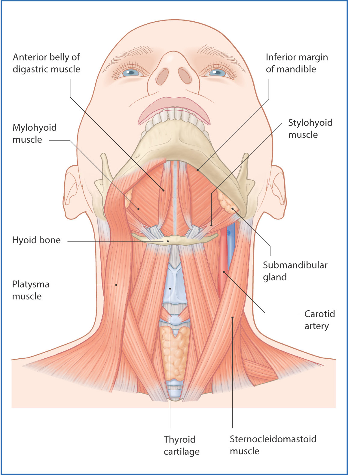
The submandibular triangle is an important anatomic landmark located underneath the body of the mandible. The majority of the anatomic space bounded by the submandibular triangle consists of the submandibular gland. The submandibular gland is the second largest salivary gland in the body (the parotid gland is the largest). In addition to housing the submandibular gland, a vast array of nerves.

PPT LIMFADENOPATI.pptx
The lymphatic system is a complex component of the immune system involved in filtering substances in the body. Lymphocytes are the integral agents involved in searching for target proteins and travel through lymph nodes, which are diffusely placed throughout the body. Lymphadenopathy is a term that refers to the swelling of lymph nodes. Lymph nodes are small glands that are responsible for.

Limfadenopati Immunodeficiency Medical
The submandibular glands are paired major salivary glands that lie in the submandibular triangle. The glands have a superficial and deep lobe separated by the mylohyoid muscle [1]. The Wharton duct, the submandibular gland's primary excretory duct, drains into the oral cavity at the sublingual caruncle. The sublingual caruncle is a papilla.

The Submandibular Gland Structure Vasculature Innervation TeachMeAnatomy
A submandibular space infection is a bacterial infection of the floor of the mouth. Bacteria can spread from an infected lower tooth to the tissue under and around the tongue. People with poor dental hygiene and people who have had a tooth pulled or a jaw fracture are at higher risk. The infection causes swelling that can block the airway.
Submandibular triangle Anatomy and clinical notes Kenhub
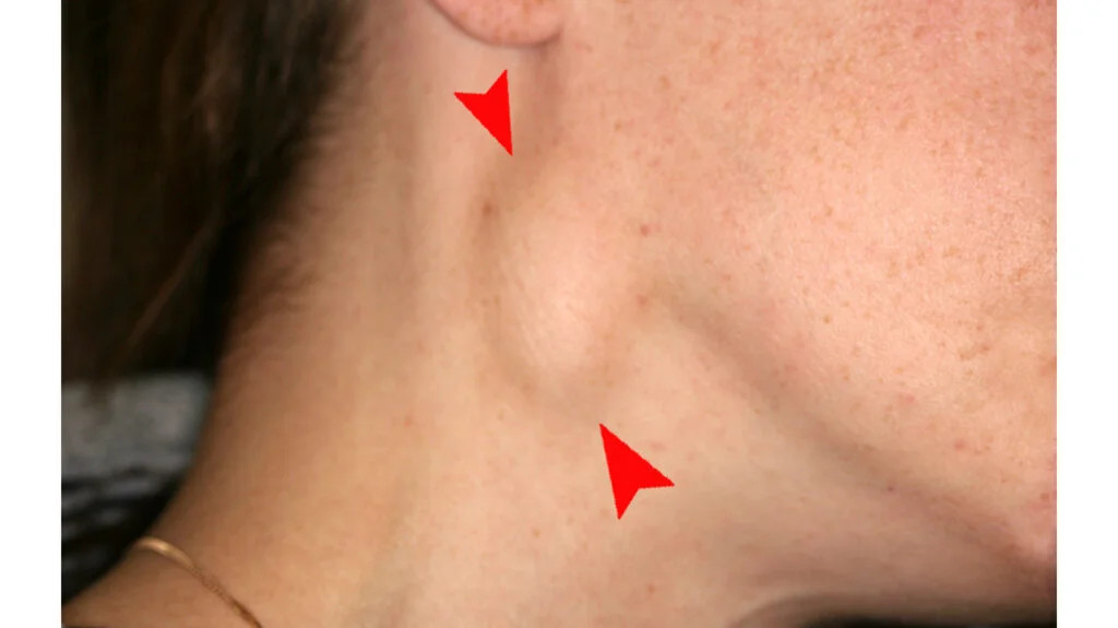
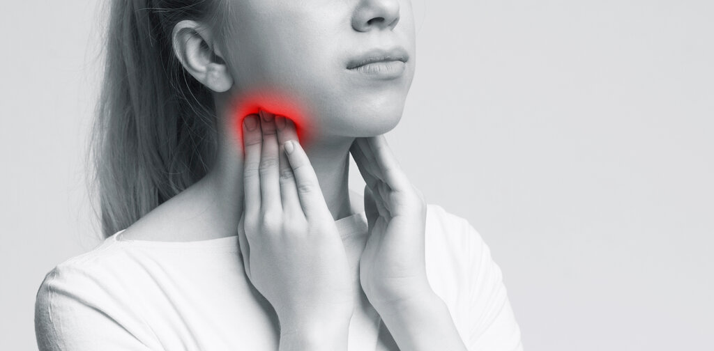
Limfadenopati submandibula merupakan istilah yang digunakan untuk menggambarkan pembesaran kelenjar getah bening yang terletak di bawah mandibula (rahang bawah). Kelenjar getah bening seharusnya tidak teraba dan merupakan rumah bagi sel-sel pertahanan tubuh dalam melawan infeksi. Pada keadaan infeksi (misal: oleh virus, bakteri, parasit.
Penyakit Limfadenopati Colli Definisi, Penyebab, Gejala, dan Tata Laksana AI Care
Submandibular lymph nodes. Superficial lymph glands and lymphatic vessels of head and neck. (Submaxillary glands labeled at center right.) The submandibular lymph nodes ( submaxillary glands in older texts), are some 3-6 lymph nodes situated at the inferior border of the ramus of mandible. [1]
Limfadenopati Lifepack.id
Analysis of the records showed the patient to be a skeletal Class II malocclusion with a deficient mandible which necessitated functional orthopedic therapy with the "Twin-Block appliance"[9,10,11] to aid in mandibular advancement enabling correction of the sagittal skeletal discrepancy initially.The case would be reassessed for further dental treatment after completion of the functional.

Figure 1 from Tuberculous Lymphadenitis and Parotitis. Semantic Scholar
Lymphoepithelial cysts are benign, slowly growing unilocular or multilocular lesions that appear in the head and neck. They are also called Branchial cyst. The head and neck sites are the salivary glands (more commonly parotid and rarely submandibular gland) and the oral cavity (usually the floor of the mouth). These cysts are usually seen in.
Linfoadenopatie laterocervicali
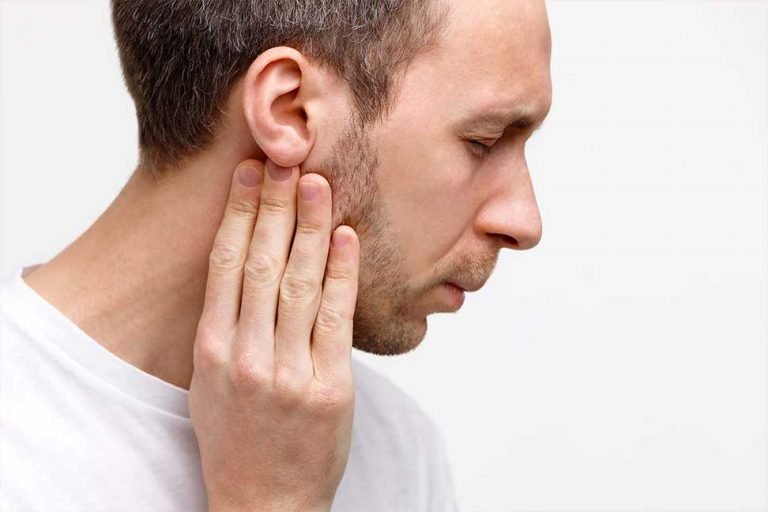
Submandibular lymphadenopathy refers to enlarged lymph nodes located beneath the mandible (lower jaw). Hot, swollen, tender, supple lymph nodes usually indicate infection and are accompanied by.
Submandibular Region Basicmedical Key
Lymphadenopathy is a common abnormal finding during the physical exam in general medical practice. Patients and physicians have varying degrees of associated anxiety with the finding of lymphadenopathy as a small number of cases can be caused by neoplasm or infections of consequence, for example, HIV or tuberculosis (TB). However, it is generally recognized that most lymphadenopathy, both.
Limfadenitis Gejala, Penyebab, Pengobatan, dll DokterSehat
Konsul pasien dengan pembesaran KGB submandibular multipel. Laki-laki, 35 tahun, mengeluhkan pembesaran KGB sejak 3 bulan lalu. Hasil USG : multiple lymphadneopathy submandibular dextra uk 1.3 x 0.9 cm dan 1 x 0.5 cm, morfologi benign. Pasien memiliki riwayat berhubungan seks dengan penderita HIV tanpa menggunakan pengaman sekitar 3 bulan lalu.

PPT LIMFADENOPATI PowerPoint Presentation, free download ID3472733
The submandibular glands are small, paired exocrine glands, each located within the submandibular (digastric) triangle of the neck. Together with the parotid and sublingual glands, they make a set of major salivary glands that are the components of the accessory digestive system. In addition, there are many minor salivary glands scattered.
:max_bytes(150000):strip_icc()/Submangland-3ef4ae5c5586481ea1669f7de9e450cc.jpg)
Submandibular Gland Anatomy, Function, and Treatment
Submandibular glands are major paired salivary glands. It is located in the submandibular triangle covered by the investing layer of deep cervical fascia. Mylohyoid muscle separates the superficial and deep lobe of the glands. Submandibular glands drain into the mouth via Wharton's duct, which courses between the sublingual gland and hyoglossus muscle; it opens through a small opening.

IT 7 Pendekatan Diagnostik Limfadenopati Limfadenitis ND Lymphatic System Biopsy
The submandibular gland produces saliva, which moistens the mouth and aids in chewing, swallowing, digestion, and helps to keep the mouth and teeth clean. Unstimulated, the submandibular glands provide the majority of saliva to the mouth. On stimulation, the parotid gland takes over, producing the majority of saliva.
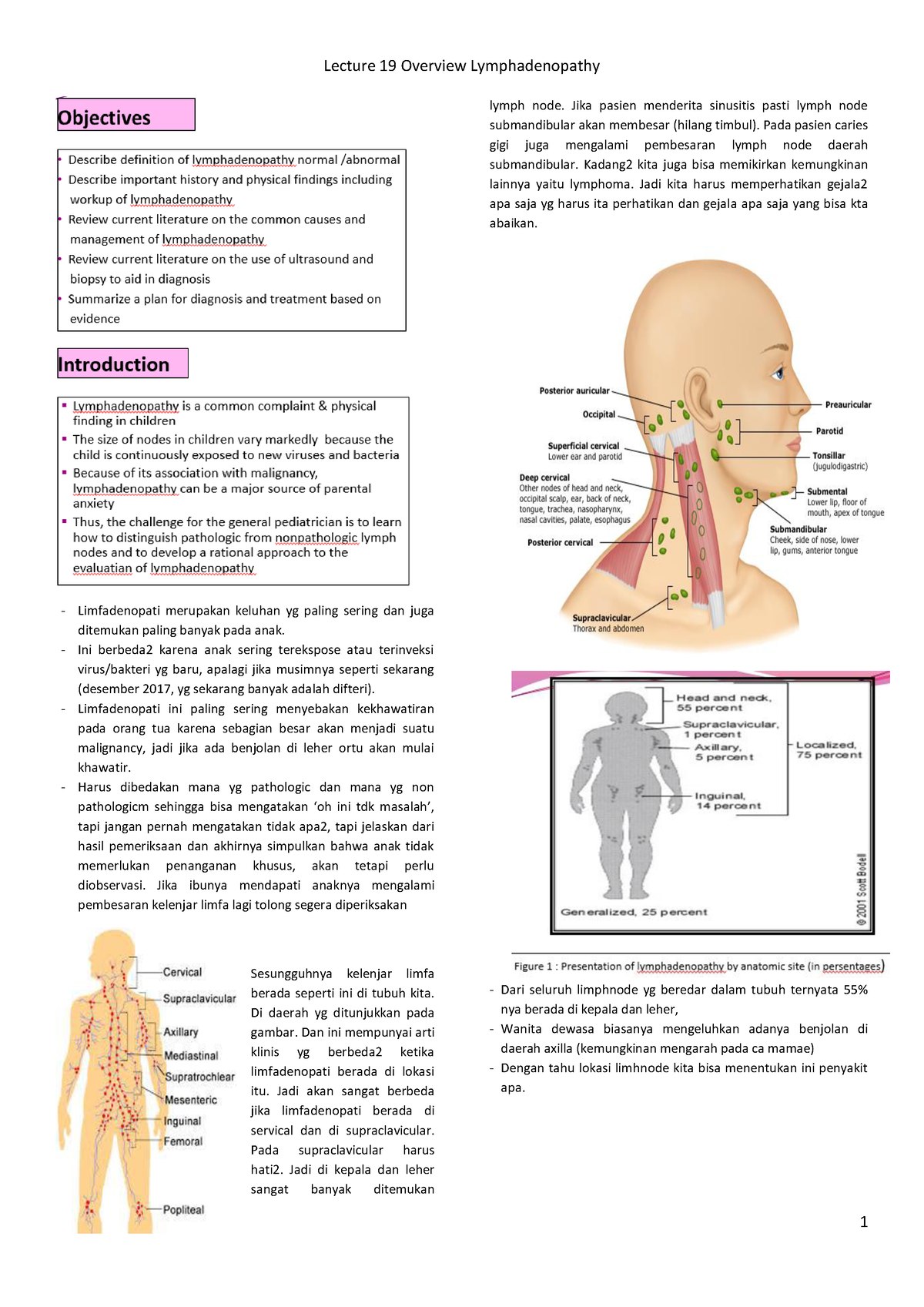
Lecture 19 Overview Lyphadenopathy Limfadenopati merupakan keluhan yg paling sering dan juga
Submandibular gland papilla. The terminal part of the submandibular (Wharton's) duct is located in the mouth floor and opens as an orifice of the submandibular duct papilla. The position of the duct and its 0.5-1.5 mm wide ostium is invariably symmetric, but quite unpredictable; consequently, submandibular duct papillae can occasionally be.
Pendekatan diagnosis limfadenopati
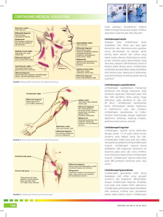
Lymphadenopathy of the right supraclavicular node is associated with cancer in the mediastinum, lungs or esophagus. The left supraclavicular (Virchow's) node receives lymphatic flow from the.