Processus xiphoideus pacs
The xiphoid process is a small bony feature of the anterior thoracic wall just inferior to the sternum corpus. Although the xiphoid process is commonly represented as a straight, fully ossified bone in educational textbooks, reports of anomalous processes flood the literature. The xiphoid process can be broad, thin, monofid, bifid, trifid.
Xiphoid Sternum
There is considerable anatomic variation in the shape of the xiphoid of the sternum:. xiphoid ending is classified as single, double, or triple.; xiphoid size varies (e.g. elongated process); xiphoid morphology (e.g. ventral or dorsal deviation, hook-like, reverse S-shape).; Clinical presentation. Elongated and ventrally-deviated xiphoid process might mimic an epigastric mass and cause pain 3,4.
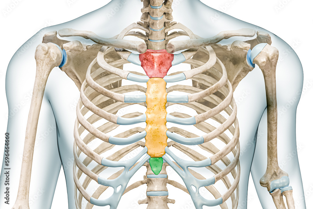
Manubrium, body and xiphoid process bones of the sternum in colors 3D rendering illustration
Summary. The xiphoid process is a small extension of bone just below the sternum. Straining and heavy lifting can damage the xiphoid process, leading to pain in the lower ribcage, breastbone, and.
Processus xiphoideus MediKarriere
Case Discussion. There is considerable anatomic variation in the shape of the xiphoid of the sternum: xiphoid ending is classified as single, double, or triple. xiphoid size varies (e.g. elongated process) xiphoid morphology (e.g. ventral or dorsal deviation, hook-like, reverse S-shape).
Xiphoid process anatomy, function & xiphoid process pain
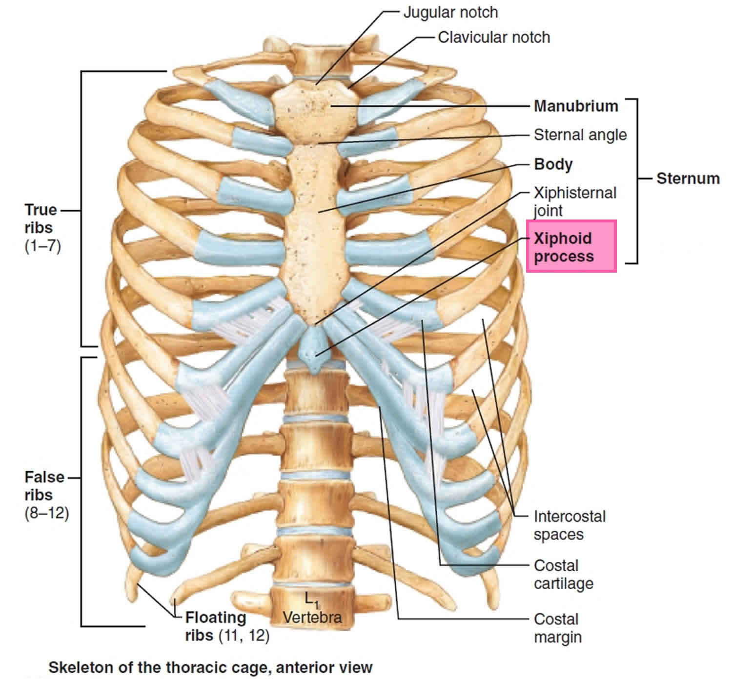
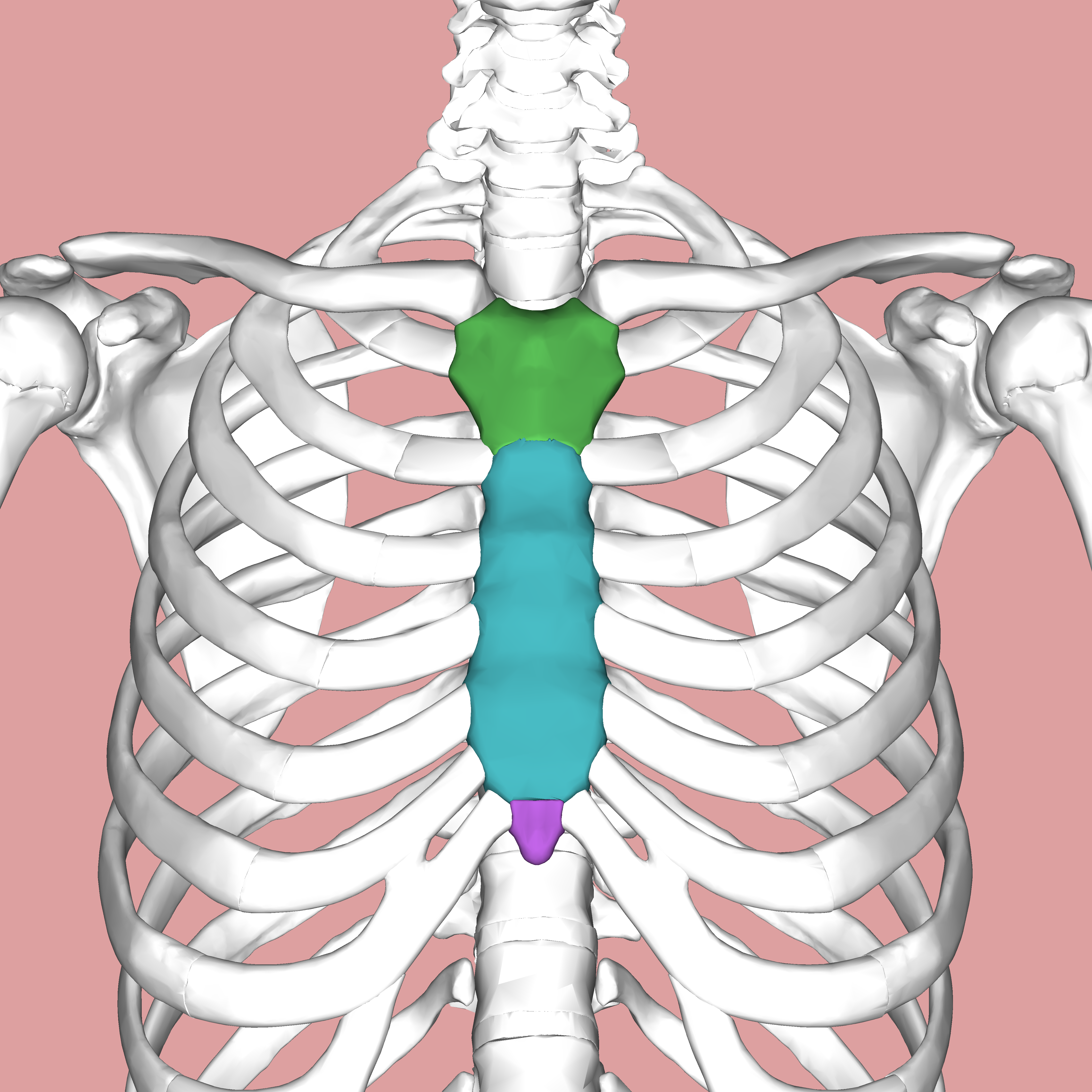
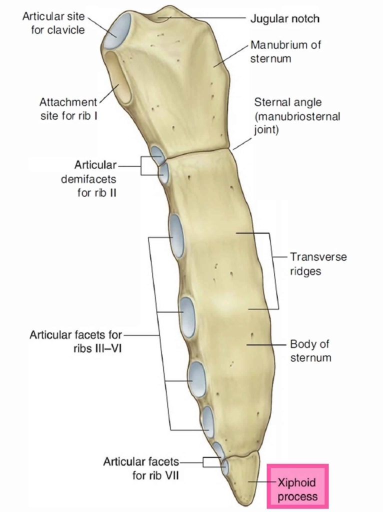
The xiphisternum (also known as the xiphoid process or simply the xiphoid) is the smallest of the three parts of the sternum ( manubrium, body or gladiolus, and xiphisternum). It arises from the inferior and posterior margin of the sternal body and projects inferiorly. It is a small cartilaginous extension of the lower sternal body, with which.

xiphoid process of the sternum
PMID: 29098125. PMCID: PMC5659327. DOI: 10.7759/cureus.1613. The xiphoid process is a small bony feature of the anterior thoracic wall just inferior to the sternum corpus. Although the xiphoid process is commonly represented as a straight, fully ossified bone in educational textbooks, reports of anomalous processes flood the literature.
xiphoid process bone
processus xiphoideus. Interactive 3D Anatomy. The BioDigital Human platform is an interactive 3D, medically accurate, virtual map of the human body—including over 8,000 individually selectable anatomical structures, 850+ simulated 3D health conditions and treatments. Explore 3D anatomy or create immersive experiences with our fully embeddable.
Xiphoid Process Pain, Lump, Removal, and More
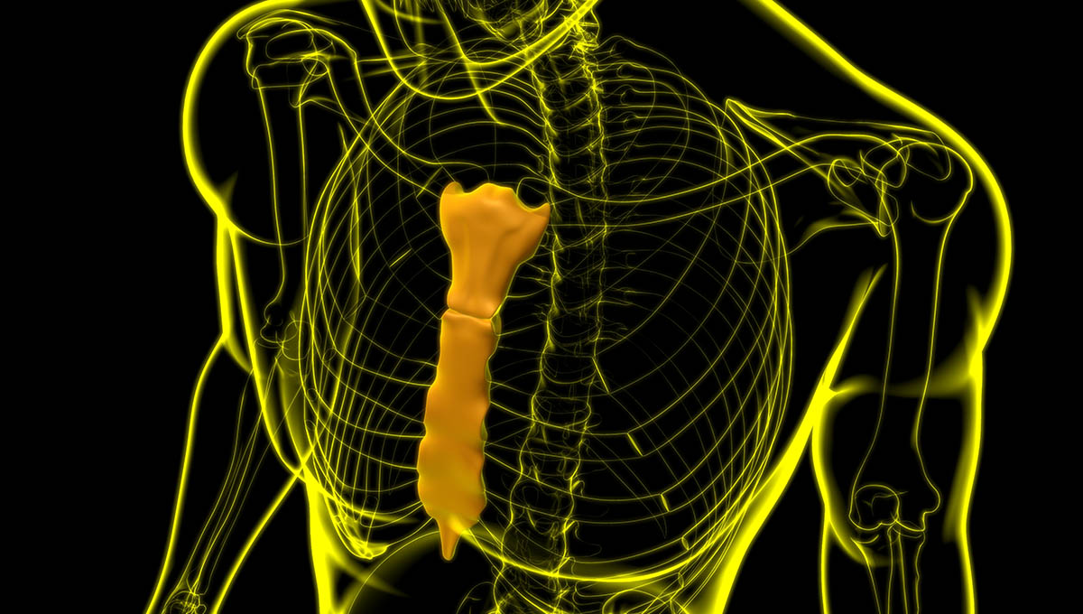
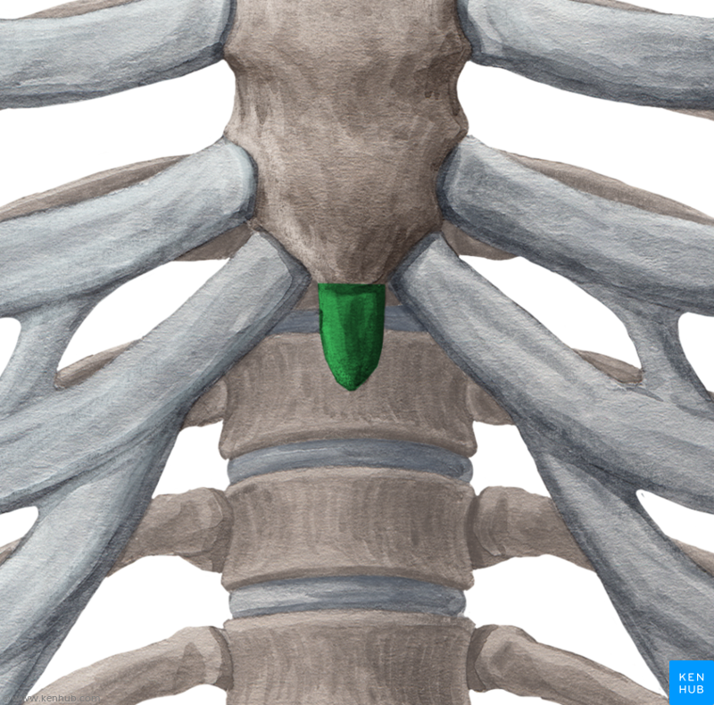
The xiphoid process (or xiphisternum, metasternum) is the third segment of the sternum, commonly referred to as the breast bone, in human anatomy. It is a small section of bone located at the base of the sternum at the 10th thoracic vertebrae. It is most commonly triangular in shape and may also features a small perforation within its structure.
xiphoid process location
The xiphoid process, also referred to as the xiphisternum or ensiform process, is a bony process that is considered the most variable and smallest part of the sternum. Morphological variations range from being pointed, bifurcated, trifurcated, broad, curved, or deflected [ 1 ]. These anatomical variations are important to consider in order to.
Xiphoid process anatomy, function & xiphoid process pain
Latin: M. psoas major.The psoas muscle forms together with the spine the dorsal boundary of the abdominal cavity. Origin at the vertebral column. The psoas muscle, together with the iliac muscle, passes through the muscular lacuna to the thigh, and acts as a hip flexor by its attachment to the lesser trochanter of the femur.
xiphoid process
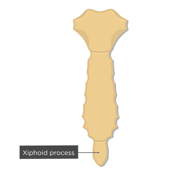
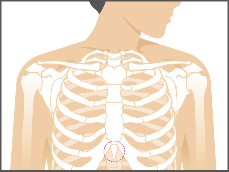
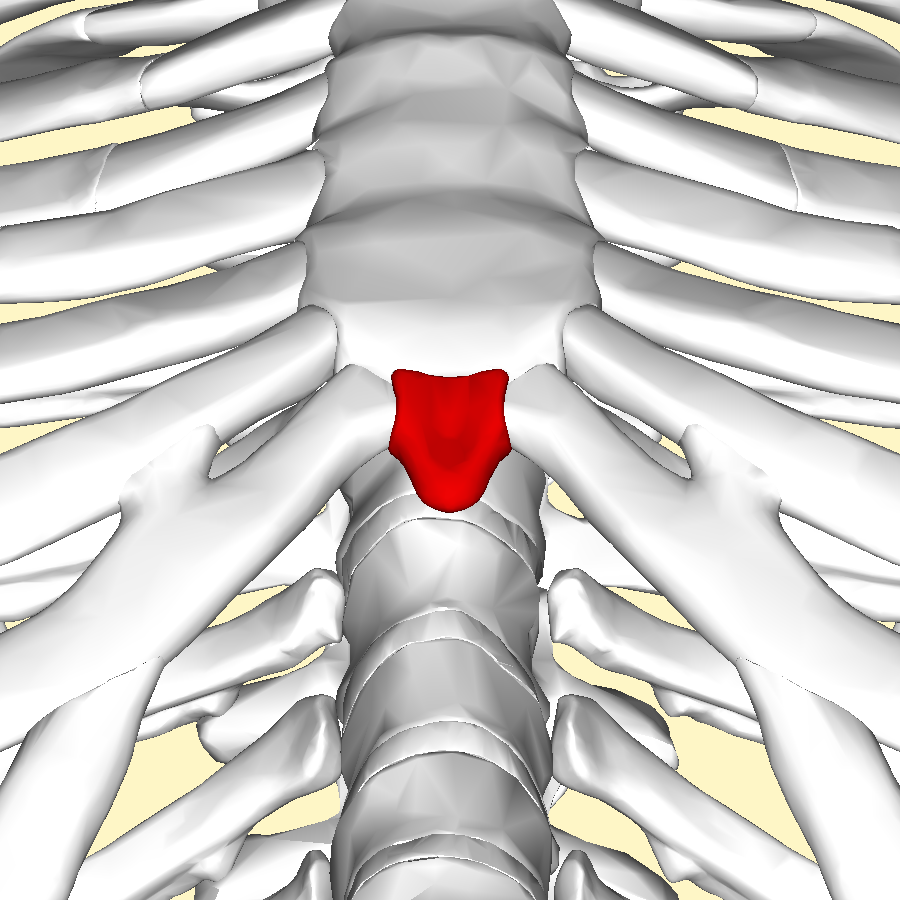
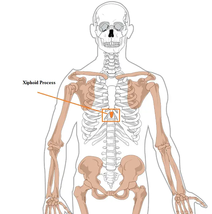
The xiphoid process is the smallest and most inferior region of the sternum, or breastbone. At birth, it is a thin, roughly triangular region of cartilage that slowly ossifies into a bone and fuses with the body of the sternum. Clinically, the xiphoid process plays an important role as a bony anatomical landmark in the trunk and may be damaged.
Processus xiphoideus pacs
A 59-year-old woman presented with a 30-year history of epigastric cutaneous protuberance. A mass was visible in the mid-portion of the epigastrium, particularly when in the supine position (Figure A). Sagittal computed tomography revealed an elongated and curved xiphoid process (Figure B). The measured length, width, and thickness of the xiphoid process were 63.2 mm (reference range, 40-50 mm.
processus xiphoideus steht vor processus xiphoideus schwellung Kuchi
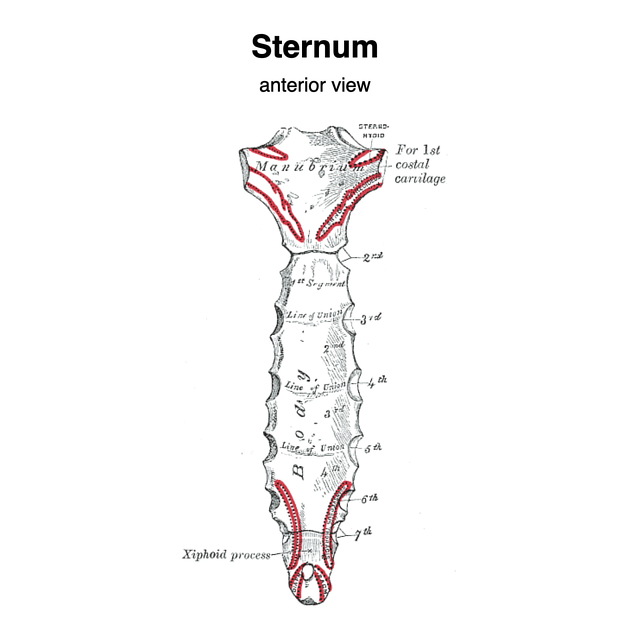
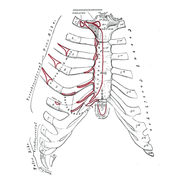
The xiphoid process is the smallest of the three pieces: it is thin and elongated, cartilaginous in structure in youth, but more or less ossified at its upper part in the adult.Surfaces.—Its anterior surface affords attachment on either side to the anterior costoxiphoid ligament and a small part of the Rectus abdominis; its posterior surface, to the posterior costoxiphoid ligament and to.

Xiphoid Process Pain, Lump, Removal, and More
The xiphoid process (/ ˈ z ɪ f ɔɪ d /), also referred to as the ensiform process, xiphisternum, or metasternum, constitutes a small cartilaginous process (extension) located in the inferior segment of the sternum, typically ossified in adult humans. Both the Greek-derived term xiphoid and its Latin equivalent, ensiform, connote a "swordlike" or "sword-shaped" morphology.
What Is The Xiphoid Process and Where Is It Located? The Healthy Apron
Processus xiphoideus Read more. Description. The xiphoid bone (xiphoid bone, xiphisternum or ensiform bone) is the thin, triangular, lower part of the sternum. It is located at the level of the tenth thoracic vertebra. Its shape can vary, where it can also appear as curved or bifid, and it contains:

Xiphoid process Pain, lump, and removal
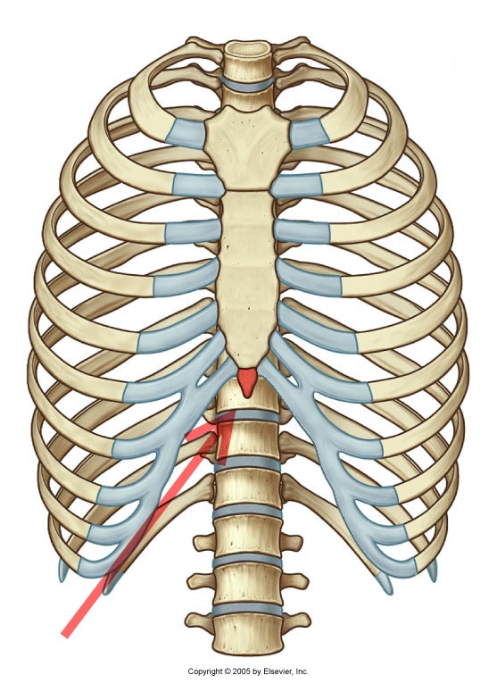
In order to examine anatomical changes in the thorax as a whole over time, a study protocol was constructed. Three different distances in mm, at three different thoracic levels (Incisura Jugularis, Angulus Sterni, and Processus Xiphoideus) were chosen and measured on axial CT images (Fig. 1).Distances at each level included a transverse distance (TVD), and two antero-posterior distances, i.e.
